MRI Abdomen
MRI Small Bowel
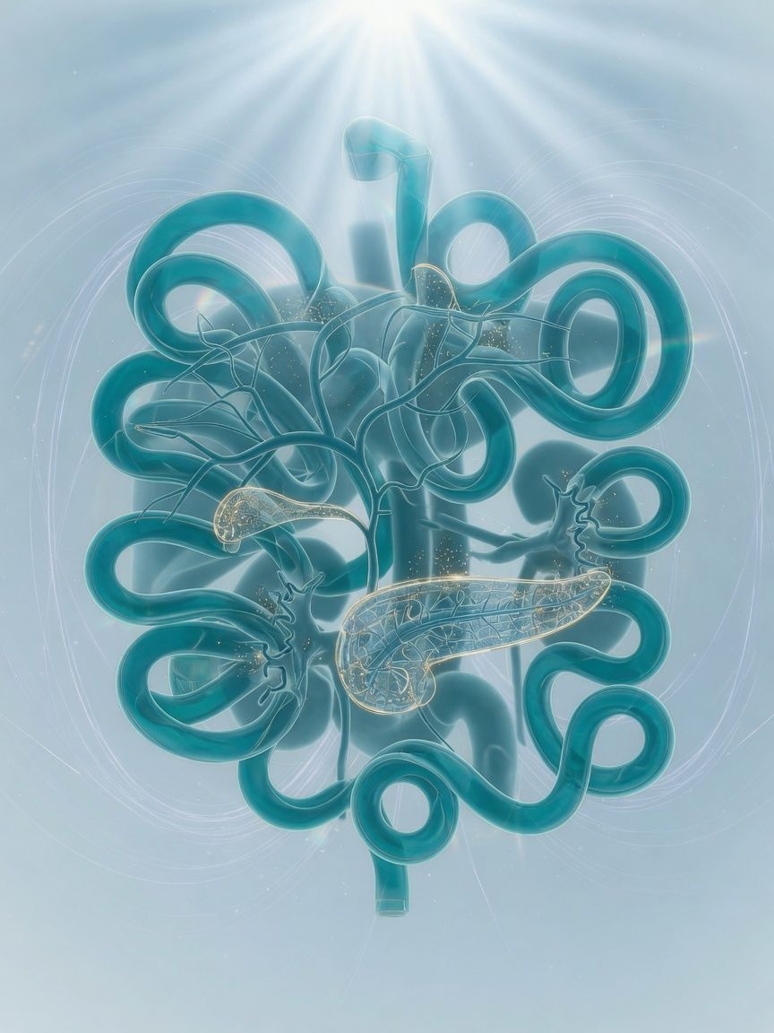
Crohn's disease: MR enterography (Sellink MRI of the small bowel) as the standard for diagnosis, activity, and follow-up – the entire small bowel including its wall, without radiation.
Top- Examination time (min)
- 30–40
- Report turnaround (days)
- 1–3
Who it is for
TopFor patients with suspected or known Crohn's disease, with unclear abdominal pain and diarrhea, or for evaluation of small bowel strictures – MRI Small Bowel (MR enterography, "Sellink MRI") in Frankfurt.
When
- Suspected Crohn's disease: detection and extent of inflamed small bowel segments, especially in the final segment (terminal ileum)
- Known Crohn's disease: assessment of inflammatory activity and treatment response without radiation exposure
- Complications such as narrowing (strictures), fistulas, or abscesses in the abdomen
- Recurrent, cramping abdominal pain with the question of an obstruction in the small bowel
- Supplement after capsule endoscopy for unclear small bowel findings or suspected tumor
When not
- Acute bowel obstruction with vomiting and a severely distended abdomen: emergency department
- Changes of the colonic mucosa (e.g., polyps, screening): colonoscopy is the appropriate examination for this
- Implant that is not MRI-compatible (e.g., pacemaker)
What the images show
Top- T2 in several planes after drinking a contrast solution: wall thickness, wall edema, strictures
- Diffusion-weighted imaging: inflammatory active bowel segments
- Fat-suppressed T1 after contrast agent: wall enhancement, fistulas, abscesses
- Motion imaging (cine): bowel motility, passage through strictures
Course
TopArrival
Registration, underlying condition and current treatment, kidney function values.
Briefing
Before the examination, about 1–1.5 liters of an oral solution are drunk over approximately 45–60 minutes; this may cause loose stools. During the examination, you will receive an antispasmodic medication (e.g., butylscopolamine) and a contrast agent through a vein, provided there is no contraindication.
Positioning
Prone or supine position, body coil over the abdomen, IV line in the arm.
Examination
Usually about 30–40 minutes in the scanner, in addition to the drinking time.
Conversation
After the examination, you will receive an initial assessment; the evaluation of the bowel segments is given in the written report.
Written report
Written report with affected segments, activity, and complications, usually after 1–3 days.
Time at the practice
Approximately 1.5 to 2 hours including the drinking phase.
Preparation
Top- Do not eat for about 4–6 hours beforehand
- Inform us in advance of glaucoma, an enlarged prostate, or cardiac arrhythmias – important for the antispasmodic medication
- Your vision may be briefly blurred after the examination; driving yourself is not advisable in that case
Report
TopAffected bowel segments with length and location, wall thickness, signs of inflammation, strictures with upstream dilatation, fistulas, abscesses, lymph nodes, mesenteric vascular signs, associated findings.
Reported by Dr. Christopher Behrens or Dr. Manuel Schloß.
Limit
Top- Superficial mucosal changes and small ulcers are not visible on MRI; endoscopy remains indispensable for these.
- Good bowel distension is a prerequisite; if the oral solution is not tolerated, the diagnostic value is limited.
Sources
Top- GuidelineMaaser C et al., ECCO/ESGAR. ECCO-ESGAR Guideline for Diagnostic Assessment in IBD Part 1: Initial diagnosis, monitoring of known IBD, detection of complications. J Crohns Colitis 2019;13:144-164. 2019.
- Paper on the next stepTaylor SA et al.. The first joint ESGAR/ESPR consensus statement on the technical performance of cross-sectional small bowel and colonic imaging. Eur Radiol 2017;27:2570-2582. 2017.
Questions
Top- How long does it take?
- Usually 30–40 minutes in the scanner, plus about 45–60 minutes of drinking time beforehand.
- When does the report arrive?
- The complete written report is usually available within 1–3 business days.
Appointment
TopWhat to bring
- Endoscopy and histology reports
- Prior images
- List of medications
- Implant card