MRI Extremities
MRI Finger
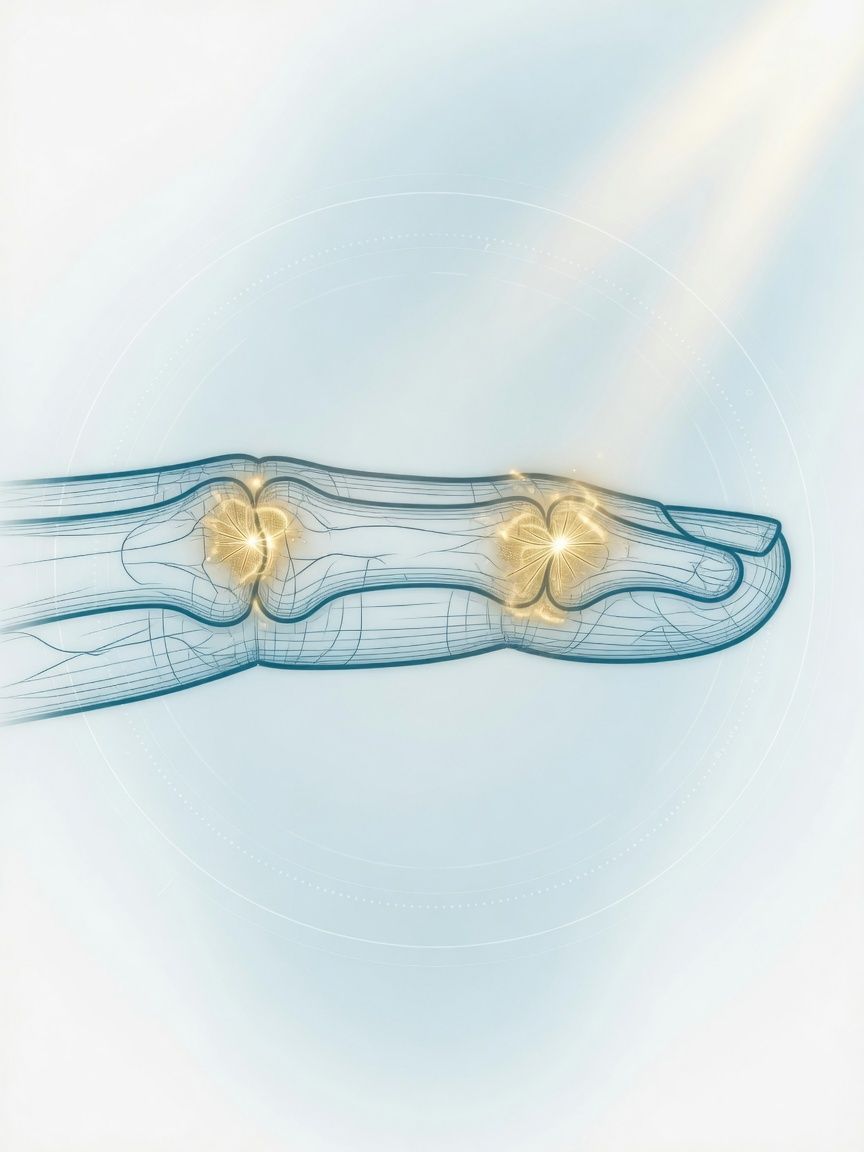
Pulleys, tendons, collateral ligaments, small soft-tissue nodules: in high resolution with MRI of the finger and thumb. Helps decide between splinting and hand surgery.
Top- Examination time (min)
- 20–30
- Report turnaround (days)
- 1–3
Who it is for
TopFor climbers, ball sports athletes, and anyone with injuries or unclear nodules on individual fingers or the thumb, when a high-resolution MRI of the finger in Frankfurt is intended to clarify the pulleys, tendons, ligaments, or nail region.
When
- Injury to the annular pulleys of the flexor tendons, typically from climbing, with pain on the palm side of the finger
- Injury to the collateral ligament of the thumb's metacarpophalangeal joint ("skier's thumb"), especially to determine whether the ligament is displaced (Stener lesion) and should be treated surgically
- Suspected flexor or extensor tendon tear or volar plate avulsion after hyperextension
- Small, very painful nodule under the fingernail (suspected glomus tumor) or other soft-tissue nodules on the finger
- Unclear bone finding in a phalanx (e.g., enchondroma) or suspected bone infection after injury or nail bed infection
When not
- Obvious finger dislocation or fracture: first X-ray and reduction
- Superficial nail bed infection without signs of deeper spread: clinical treatment
- Implant that is not MRI-compatible (e.g., pacemaker)
What the images show
Top- Very thin slices with a small field of view: annular pulleys, collateral ligaments, volar plate
- Sagittal slices along the finger: distance between the flexor tendon and the bone (sign of a pulley rupture)
- T1 and fat-suppressed sequences: bone marrow, glomus tumor, enchondroma
- Contrast agent for suspected glomus tumor, nodules, or inflammation
Course
TopArrival
Registration, affected finger, mechanism of injury (e.g., climbing grip, ball injury).
Briefing
The finger is secured in a small coil; please do not move. Contrast agent only for nodules or inflammation.
Positioning
Depending on the scanner, prone position with the arm extended forward or supine position, finger extended on a splint.
Examination
Usually about 20–30 minutes.
Conversation
We show you the images and explain the findings after the examination.
Written report
Written report usually after 1–3 days.
Time at the practice
Approximately 45 minutes to 1 hour.
Preparation
Top- Remove all rings
- No need to fast
- Remove splint or tape after consultation on site
Report
TopAnnular pulleys stating the affected pulley, flexor and extensor tendons, collateral ligaments, volar plate, bones of the phalanges, soft-tissue or nail bed nodules.
Reported by Dr. Christopher Behrens or Dr. Manuel Schloß.
Limit
Top- Because the structures are very small, keeping still is crucial; even slight movement reduces the diagnostic value.
- For pulley injuries, dynamic ultrasound is an equivalent and often faster alternative; MRI complements it in case of unclear findings or multiple injuries.
Sources
Top- Paper on the next stepRose S et al.. Patient-Friendly Summary of the ACR Appropriateness Criteria: Acute Hand and Wrist Trauma. J Am Coll Radiol 2021;18:e3. 2021.
- Paper on the next stepYalcindag SE et al.. Patient-Friendly Summary of the ACR Appropriateness Criteria: Soft-Tissue Masses. J Am Coll Radiol 2023;20:e7. 2023.
Questions
Top- How long does it take?
- Usually 20–30 minutes.
- When does the report arrive?
- The complete written report is usually available within 1–3 business days.
Appointment
TopWhat to bring
- X-ray images of the finger
- Prior hand surgery reports
- Implant card