MRI Extremities
MRI Foot
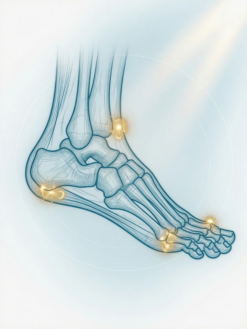
Stress fracture, nerve nodule between the toes, plantar fasciitis, bone infection in diabetes: MRI of the foot as the basis for rest from loading, insoles, or treatment.
Top- Examination time (min)
- 25–35
- Report turnaround (days)
- 1–3
Who it is for
TopFor people with forefoot, midfoot, or heel pain, with suspected stress fracture, Morton's neuroma, or inflammation – including in diabetes – that is to be clarified more precisely with an MRI of the foot in Frankfurt.
When
- Forefoot pain with numbness between the toes: suspected Morton's neuroma or bursitis between the metatarsal heads
- Exercise-related pain in the midfoot in runners or after hiking ("march fracture"): stress fracture, often before it is visible on X-ray
- Persistent heel pain with the question of inflammation or tear of the plantar fascia or a stress fracture of the heel bone
- Diabetic foot syndrome: question of bone infection (osteomyelitis), abscess, or Charcot foot
- Pain under a metatarsal head with the question of a plantar plate injury or impaired blood supply (e.g., Köhler disease)
When not
- Hallux valgus or osteoarthritis of the big toe joint without an additional question: assessment is made on weight-bearing X-ray
- Recent injury with an obvious fracture on X-ray
- Implant that is not MRI-compatible (e.g., pacemaker)
What the images show
Top- Fat-suppressed sequences (STIR, PD-fs): bone marrow edema in stress fracture, osteomyelitis, or Charcot foot
- T1: fracture line, replacement of fatty marrow in bone infection – important for distinguishing it from edema alone
- Coronal and axial slices of the forefoot, possibly in prone position: Morton's neuroma, plantar plate
- Contrast agent for suspected infection (abscess, fistula) or soft-tissue nodules
Course
TopArrival
Registration, side and exact location (heel, midfoot, forefoot, toe); in diabetes, information about wounds.
Briefing
The foot is secured in the coil; for questions of infection or nodules, contrast agent through a vein.
Positioning
Supine position, foot in the foot coil; for the forefoot, sometimes prone position with the foot extended.
Examination
Usually about 25–35 minutes.
Conversation
After the examination, we go through the images with you.
Written report
Written report usually after 1–3 days.
Time at the practice
Approximately 45 minutes to 1 hour.
Preparation
Top- No need to fast
- Wound dressings can usually be left in place; please mention them at registration
- Wear socks without metal fibers
Report
TopTarsal and metatarsal bones with edema, fracture lines, and fatty marrow, joints, plantar fascia, plantar plates, intermetatarsal spaces, tendons, soft tissues with abscess or fistula.
Reported by Dr. Christopher Behrens or Dr. Manuel Schloß.
Limit
Top- In the diabetic foot, distinguishing between Charcot foot and bone infection can be difficult even on MRI; if in doubt, a tissue sample or bone biopsy remains decisive.
- Small Morton's neuromas under about 5 mm cannot always be reliably delineated; the clinical examination remains important.
Sources
Top- Paper on the next stepSaboo YS et al.. Patient-Friendly Summary of the ACR Appropriateness Criteria: Chronic Foot Pain. J Am Coll Radiol 2026;23:1462. 2026.
- GuidelineBaker JC et al., ACR. ACR Appropriateness Criteria Suspected Osteomyelitis of the Foot in Patients With Diabetes Mellitus: 2025 Update. J Am Coll Radiol 2025;22:S713-S737. 2025.
Questions
Top- How long does it take?
- Usually 25–35 minutes.
- When does the report arrive?
- The complete written report is usually available within 1–3 business days.
Appointment
TopWhat to bring
- X-ray images of the foot
- Wound reports and laboratory values in diabetes
- Implant card