MRI Joints
MRI Wrist
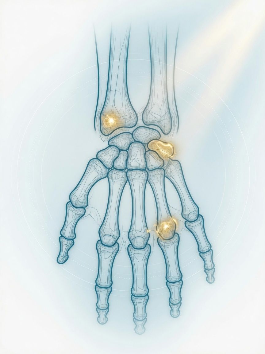
Occult fractures, ligament and TFCC injuries, early inflammation – not yet visible on X-ray. MRI of the wrist as the basis for deciding whether to immobilize, observe, or operate.
Top- Examination time (min)
- 20–30
- Report turnaround (days)
- 1–3
Who it is for
TopFor patients with wrist pain after a fall, with unclear chronic symptoms on the thumb or little finger side, or with suspected inflammatory joint disease, who need an MRI of the wrist in Frankfurt for clarification.
When
- Suspected scaphoid fracture (not visible on X-ray) after a fall on the outstretched hand
- Pain on the little finger side with the question of an injury to the triangular fibrocartilage complex (TFCC)
- Suspected ligament injury between the carpal bones (e.g., SL ligament) with instability or loss of strength
- Unclear wrist pain with the question of impaired blood supply to the lunate (Kienböck's disease) or a ganglion
- Early diagnosis or follow-up of inflammatory joint disease (e.g., rheumatoid arthritis): synovitis, bone marrow edema, erosions
When not
- Acute fracture with malalignment that is clinically and radiographically obvious: treatment takes priority
- Uncomplicated carpal tunnel syndrome: the diagnosis is made clinically and neurologically; MRI is only useful in atypical cases or if a mass is suspected
- Implant that is not MRI-compatible (e.g., pacemaker)
What the images show
Top- High-resolution coronal slices (PD with fat suppression): TFCC, ligaments, cartilage
- Coronal T1: fracture line in the scaphoid, bone marrow, impaired blood supply
- STIR / fat-suppressed sequences: bone marrow edema as a sign of recent injury or inflammation
- Contrast agent for questions of synovitis (rheumatic disease), viability of the scaphoid after fracture, or tumor
Course
TopArrival
Registration, side, time of injury or onset of symptoms.
Briefing
The hand is secured in a small coil and must remain still. Contrast agent only if the clinical question requires it.
Positioning
Depending on the scanner, prone position with the arm above the head or supine position with the hand beside the body.
Examination
Usually about 20–30 minutes.
Conversation
After the examination, the images are presented to you and the findings are discussed.
Written report
Written report usually after 1–3 days.
Time at the practice
Approximately 45 minutes to 1 hour; somewhat longer with contrast agent.
Preparation
Top- Remove rings, wristwatch, and bracelets
- No need to fast
- Discuss any cast or splint with the practice team beforehand
Report
TopCarpal bones with fracture lines and bone marrow edema, triangular fibrocartilage complex (TFCC), ligaments between the carpal bones, cartilage, tendons and tendon sheaths, ganglia, synovitis.
Reported by Dr. Christopher Behrens or Dr. Manuel Schloß.
Limit
Top- Very fine ligament tears are not always unambiguous despite high-resolution technique; in individual cases, arthroscopy remains the reference standard.
- CT is often more informative for assessing bony healing after a scaphoid fracture.
Sources
Top- Paper on the next stepMohan J et al.. Patient-Friendly Summary of the ACR Appropriateness Criteria: Chronic Hand and Wrist Pain: 2024 Update. J Am Coll Radiol 2025;22:147. 2025.
- Paper on the next stepRose S et al.. Patient-Friendly Summary of the ACR Appropriateness Criteria: Acute Hand and Wrist Trauma. J Am Coll Radiol 2021;18:e3. 2021.
Questions
Top- How long does it take?
- Usually 20–30 minutes.
- When does the report arrive?
- The complete written report is usually available within 1–3 business days.
Appointment
TopWhat to bring
- X-ray images, if available
- Prior rheumatology reports
- Implant card
- Referral with clinical question