Special
MRI Temporomandibular Joint
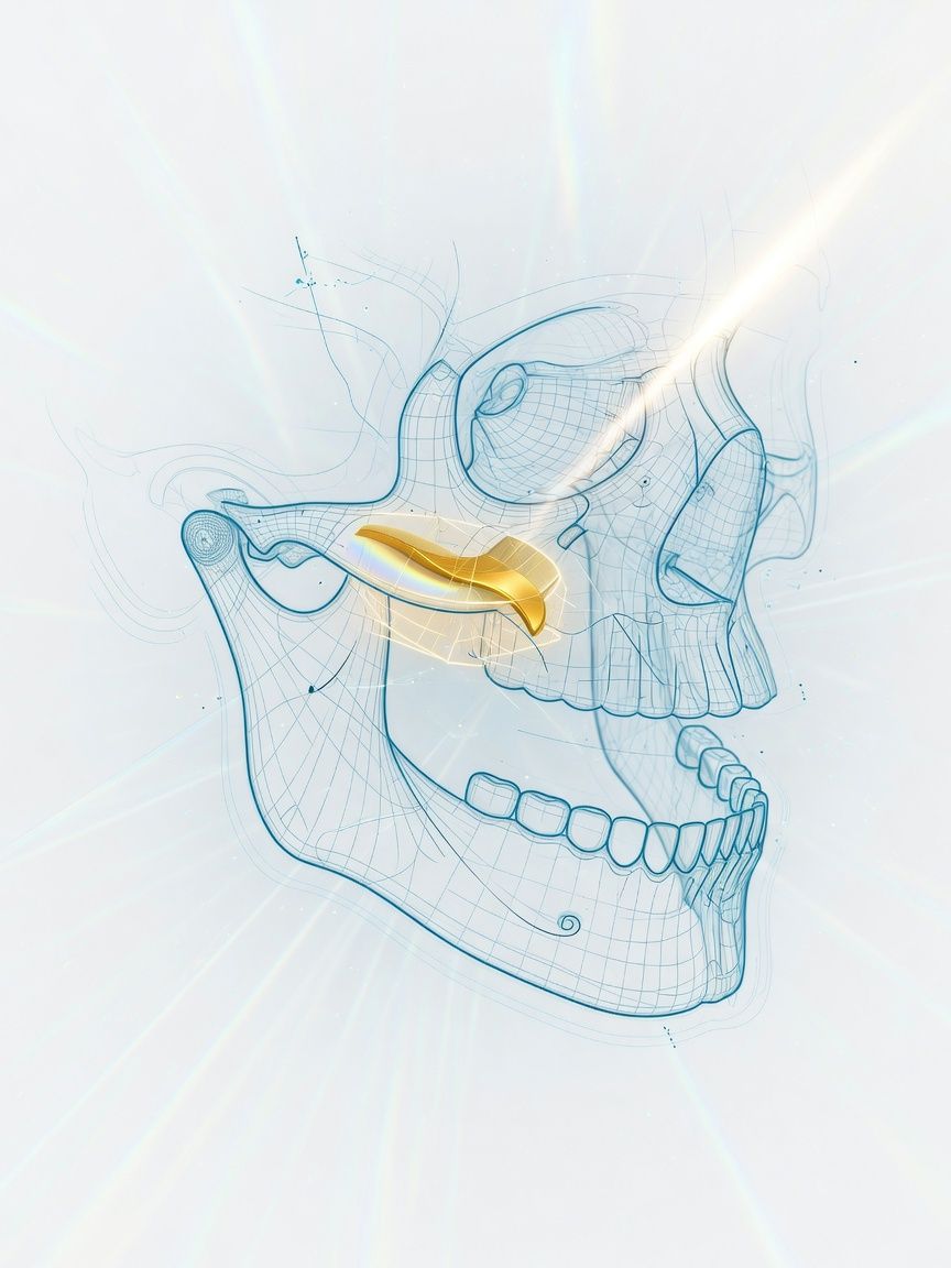
Two positions, mouth open and closed: MRI of the temporomandibular joint shows the joint and neighboring structures in high resolution – and thus also its function, for example that of the articular disc.
Top- Examination time (min)
- 25–35
- Report turnaround (days)
- 1–3
Who it is for
TopFor patients with temporomandibular joint symptoms such as clicking, pain, or restricted mouth opening, with suspected disc displacement, osteoarthritis, or joint inflammation after a dental or orthodontic examination – MRI Temporomandibular Joint in Frankfurt.
When
- Suspected disc displacement with or without reduction
- Unclear temporomandibular joint symptoms (restricted mouth opening, "clicking," and pain)
- Suspected osteoarthritis or synovitis (arthritis) of the temporomandibular joint
When not
- For questions about: dental status, tooth roots, inflammation of the maxillary sinuses
- Acute disc dislocation that must first be reduced clinically
- Symptoms without an examination by a dentist or orthodontist
What the images show
Top- PD or T2 open and closed, disc position in both positions
- T1 for imaging the bone and bone marrow
- Contrast agent only if inflammation is specifically suspected; as a rule, the examination is performed without it
Course
TopArrival
Registration, clarification of formal questions and type/duration of symptoms
Briefing
The examination is performed with the mouth closed and open; a bite block is provided.
Positioning
Supine position, head coil, jaw at the isocenter.
Examination
Usually about 25–35 minutes, because both positions are imaged.
Conversation
The images are presented to you after the examination, and the findings are discussed calmly with you.
Written report
Written report with disc position in both positions, usually after 1–3 days
Time at the practice
Approximately 45 minutes to 1 hour
Preparation
Top- Bring the dental report
- Remove removable dentures if they contain metal
Report
TopDisc position closed and open, reduction, effusion, condyle, side.
How to read it
The report describes the position of the articular disc with the mouth closed and open, and whether it slides back into its normal position on opening (reduction). Displacement with reduction without pain or functional impairment is common and on its own usually does not require splint therapy; the decision on treatment is made by the dental examination together with the imaging findings.
Reported by Dr. Christopher Behrens or Dr. Manuel Schloß.
Limit
Top- MRI shows teeth, tooth roots, and fine bony details only to a limited extent; X-ray or cone beam CT (CBCT) is better suited for this.
- Displacement of the articular disc is also found in many people without symptoms; the imaging findings therefore do not replace the clinical functional examination.
Sources
Top- GuidelineDGFDT und DGZMK (AWMF). S2k guideline on occlusal splints for the treatment of craniomandibular dysfunction and for preprosthetic therapy. AWMF 083-051, Stand Februar 2024, Abschnitt zur Behandlungsbedürftigkeit von Diskusverlagerungen. 2024.
- Paper on the next stepSchiffman E et al., International RDC/TMD Consortium Network. Diagnostic Criteria for Temporomandibular Disorders (DC/TMD) for Clinical and Research Applications. J Oral Facial Pain Headache 2014;28:6-27. 2014.
Questions
Top- How long does it take?
- Usually 25–35 minutes, two mouth positions.
- Is contrast needed?
- Usually not. Only if joint inflammation is specifically suspected.
- Do I need a referral?
- No statutory health insurance referral is required for billing of privately insured and self-pay patients. A request from a dentist, orthodontist, or oral and maxillofacial surgeon with clinical functional findings is helpful.
- When does the report arrive?
- The complete written report is usually available within 1–3 business days.
Appointment
TopWhat to bring
- Request from a dentist or orthodontist
- Side of the symptoms