Chest
MRI Thorax
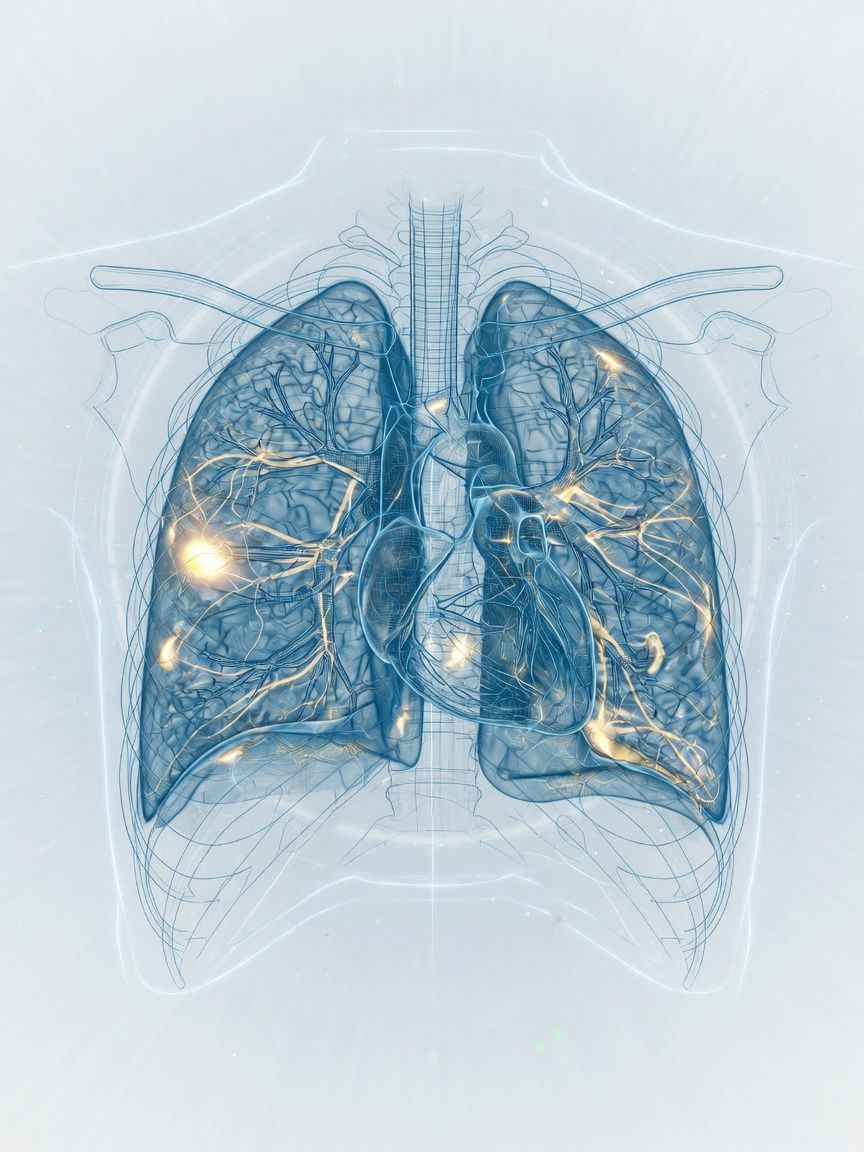
Organs and vessels of the chest, and to a limited extent the lungs – without radiation and noninvasive. Chest MRI helps with inflammation, enlarged lymph nodes, and the effects of trauma after a rib contusion.
Top- Examination time (min)
- 25–40
- Report turnaround (days)
- 1–3
Who it is for
TopFor patients with an unclear mass in the mediastinum, thymic or lymph node findings, tumors near the chest wall or spine, a soft tissue infection of the chest, or a suspected diaphragmatic hernia – MRI Thorax in Frankfurt, often as a complement to CT.
When
- Questions about a mediastinal mass (e.g., esophageal tumor), thymic disease, tumors near the spine, or abnormally enlarged lymph nodes (for example, suspected lymphoma)
- In some cases also for a suspected lung tumor or for staging of known tumors.
- Suspected infection of the soft tissues of the chest
- Suspected diaphragmatic hernia.
When not
- Question about assessing a small pulmonary nodule (under 5 mm)
- Chronic changes of the lung tissue (e.g., pulmonary fibrosis or emphysema)
- Acute shortness of breath of unclear cause
- Suspected diseases of the heart muscle or coronary arteries
What the images show
Top- T2 and T1 for the mediastinum and chest wall
- Contrast agent for tumor and pleural questions
- Respiratory triggering or breath-hold phases to counter motion
Course
TopArrival
Registration, assessment of ability to follow breathing instructions.
Briefing
Information about the type of examination (including breathing instructions); if applicable, also about intravenous contrast agent if needed
Positioning
Supine position, body coil.
Examination
Usually about 25–40 minutes.
Conversation
The images are presented to you after the examination, and the findings are discussed calmly with you
Written report
Written report, usually 1–3 days after the examination.
Time at the practice
Approximately 45 minutes to 1 hour.
Preparation
Top- Bring your previous CT report; MRI can build on it or usefully complement it
- Calm breathing; no fasting required
Report
TopCompartment, relation to vessels and chest wall, comparison with CT.
Reported by Dr. Christopher Behrens or Dr. Manuel Schloß.
Limit
Top- CT shows small pulmonary nodules and chronic changes of the lung tissue such as fibrosis or emphysema more precisely.
- Respiratory and cardiac motion can reduce image sharpness; the examination therefore requires repeated breath-holding on command.
Sources
Top- GuidelineAckman JB et al., American College of Radiology. ACR Appropriateness Criteria® Imaging of Mediastinal Masses. J Am Coll Radiol 2021;18:S37-S51. 2021.
- Paper on the next stepHatabu H et al., Fleischner Society. Expanding Applications of Pulmonary MRI in the Clinical Evaluation of Lung Disorders: Fleischner Society Position Paper. Radiology 2020;297:286-301. 2020.
- Paper on the next stepCarter BW et al., International Thymic Malignancy Interest Group (ITMIG). A Modern Definition of Mediastinal Compartments. J Thorac Oncol 2014;9:S97-S101. 2014.
Questions
Top- How long does it take?
- Usually 25–40 minutes, with breathing instructions.
- When does the report arrive?
- The complete written report is usually available within 1–3 business days.
Appointment
TopWhat to bring
- CT and/or X-ray images, if available
- If applicable, a referral with exact information on the suspected condition.
- Implant card